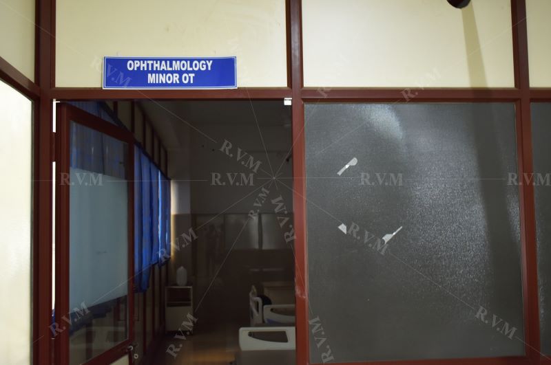
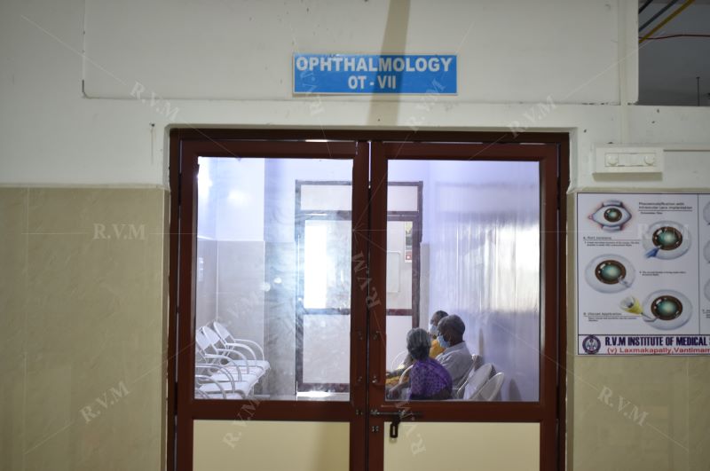
Department of Ophthalmology
Our Specialties:
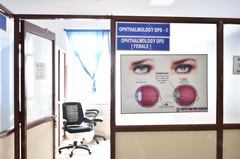
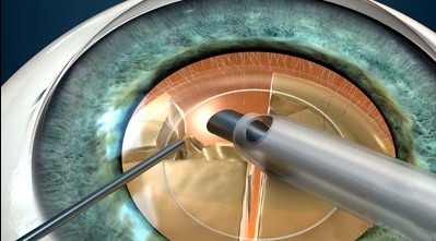
Phaco Training:
We offer the best Phaco Training course. This step by step course will give the surgeons confidence to perform surgeries independently after the completion of the course.
Advanced instrumentation and Experienced Faculty are available in our hospital. The aim of the course is to make the trainees confident enough to handle their own procedure at the end of the course.
Duration of the course – 1 Month
The candidate will be given many independent Phaco cases in 1 Month. The candidate will be taught on the easy way with different techniques for different types of cataracts. The candidates can also see the post operative patients to assess the success of their operations.
The candidate should carry a Log Book where they can record all their cases, complications, etc.,
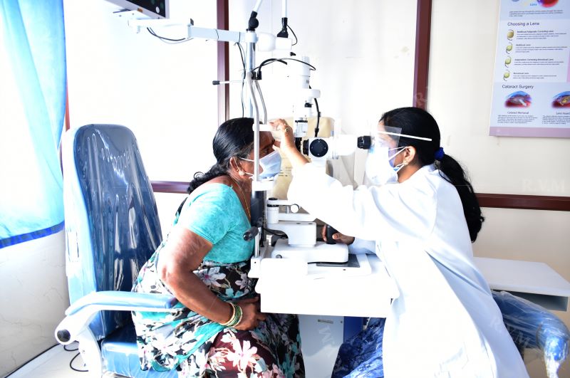
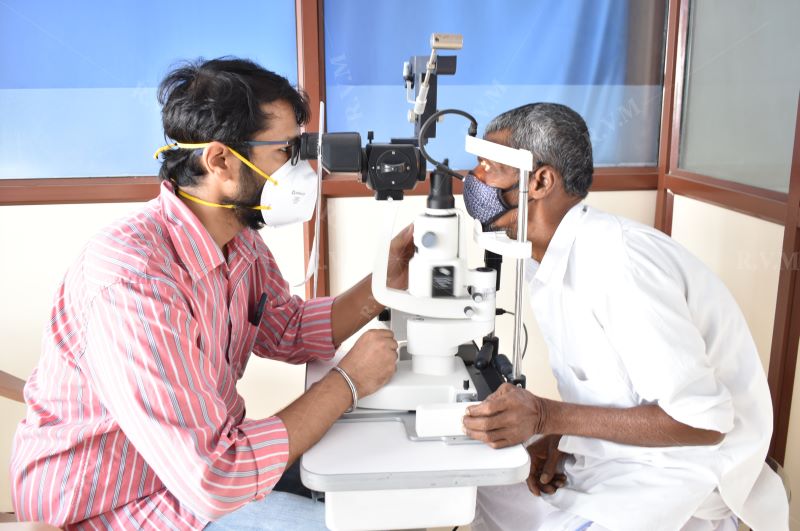
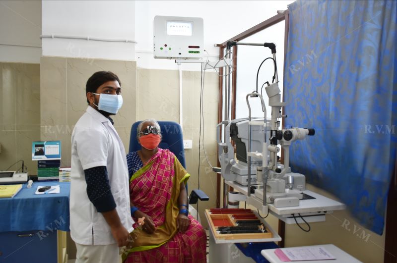
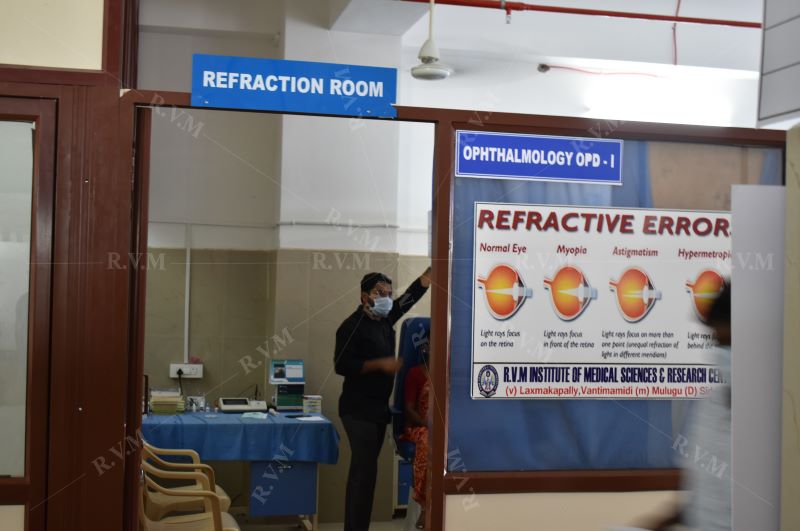
Eye Testing
We offer:
- Humphery FDT visual fields testing
- Glaucoma testing
- Children's vision
- Cataract assessment
- Contact lens fitting
Kids Vision
wondering why is vision so important?
That's because children's learning is done visually and all the hard work of reading, writing, blackboard work and computers cannot be met. It stands to reason that having a clear vision is necessary for every child. Most parents are of the opinion that it is OK so long as the child can see the blackboard.
But unfortunately, it is not so. The blackboard may still look clear and sharp to child who is having problems with his/her eyes because it has always looked that way. A simple distance vision check for the blackboard does not detect other problems which affect reading and focus.
As parents and caregivers...
If symptoms of visual stress are present parents, caregivers, and teachers should consider undetected visual problems, a contributing factor if child's learning is not keeping pace with other indicators of ability and intelligence.
Symptoms indicating vision problems:
- Headaches
- Rubbing burning or itchy eyes
- Poor concentration
- Losing place when copying
- Reversing letters or omitting short words
- Squinting frowning
- Closing one eye to look at things
- Sitting close to television
- Holding a book close to their face
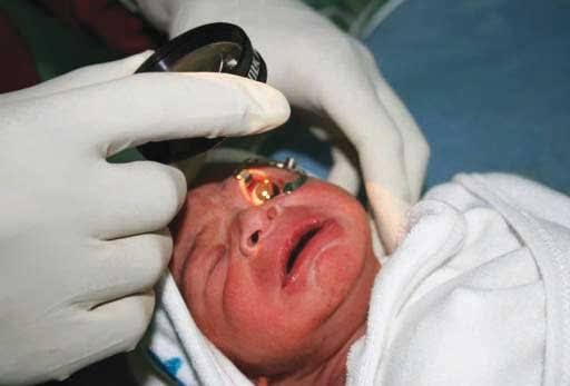
First eye examination for child
An infant child should be taken for his/her first eye exam when it is 6 months old. The next exam should be scheduled at age 3, and then again prior to your child entering first grade. A good eye doctor can test many aspects of function at this young age and quickly effect changes with intervention.
Color Vision Test
Color Vision
Objects reflect different wavelengths of light consequently producing the sensation of color and the appreciation of color is a function of the cones, three types of which are maximally sensitive to either blue, green, or red. The most common test of color vision is the Ishihara pseudo-iso-chromatic color plate exam, which is a series of test plates where matrix of colored dots is arranged with number visible on the page. The numbers are visible to individuals with normal color vision but are confused or invisible to those individuals who are red/green color deficient.
Prevention
Inherited color vision deficiency cannot be prevented. Since many learning materials are color coded, it is important to diagnose color vision deficiency early in life, which is why we recommend a comprehensive optometric examination before a child begins school.
Treatment
Congenital (inherited) color vision deficiency cannot be treated or cured, but measures can be taken, to compensate for it. Some people develop their own system of recognizing colors by their brightness and location. Acquired color vision deficiency requires treatment of the underlying cause. In many cases, normal color perception returns when the underlying condition has been resolved.
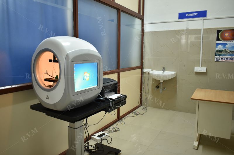
Visual Field Testing
Most people often incorrectly assume that they can see their hand waving off in the periphery, and that their peripheral vision is fine. This would include glaucoma, strokes, and brain tumor. The visual filed is the area of space that can be seen at the same time. The area in space that may be visualized by the eye is known as the visual filed and plotting of the visual field is important for many particularly disorders of the optic nerve and brain.
We have the largest computerized visual filed analyzer, the Humphrey Matrix that is able to detect common and subtle peripheral vision deficits, in which the device systematically plots the field of vision using threshold testing.
Glaucoma Testing
The following methodologies are in practice:
- Intraocular pressure is measured and the patient is under 24 hrs observation Schiotz & Applanation tonometers are employed.
- Gonioscopy is used to detect whether it is closed or open angled.
- Perimetry is used for visual fields with Goldman's & Automatic (Humprey's).
- Optic Disc cupping is detected & C:D ration is determined.
- Fundus changes are clinically observed and assessed.
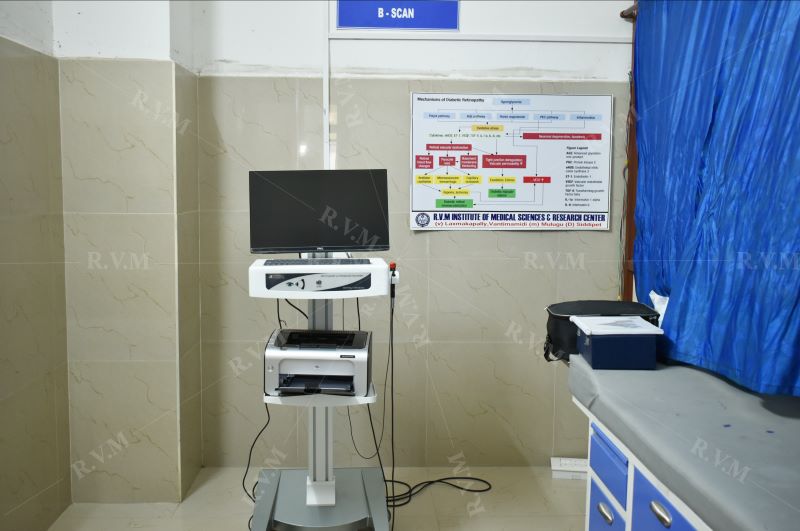
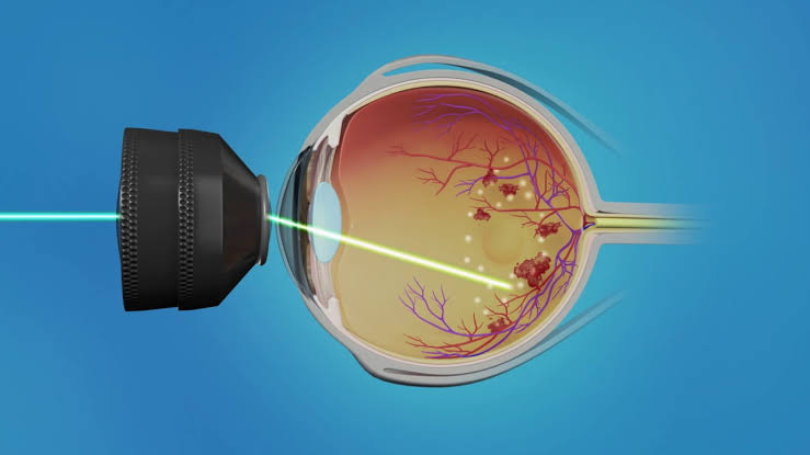
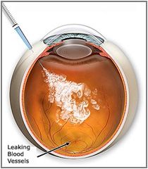
Diabetes Retinopathy Evaluation:
Fundus changes observed and treated accordingly.